

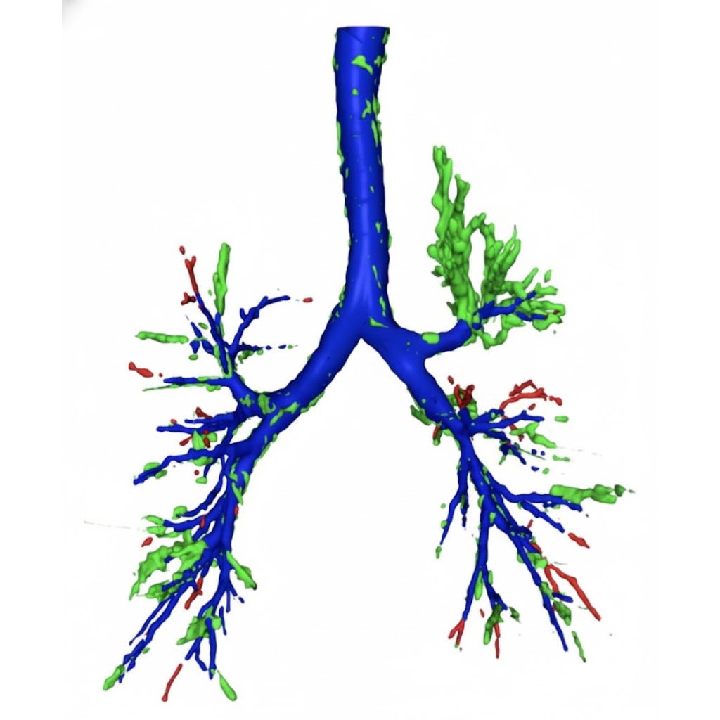
3D reconstruction of the airways generated from a CT scan using AI
Image source: University of Southampton
To address this challenge, the research team created a deep learning model. It combines a high-precision airway mapping technique (MedpSeg) with a neural network that analyses CT images for hidden signs of foreign bodies. The model was trained and tested using three independent patient groups, consisting of over 400 patients, in collaboration with hospitals in China.
To put the model to the test, researchers compared its performance to that of three expert radiologists, each with over ten years of clinical experience. The task was to examine 70 CT scans, 14 of which were cases of radiolucent FBA, confirmed by bronchoscopy.
When the radiologists detected a case of radiolucent FBA, they did so with total precision – there were no false positives. In comparison, the AI model did so with 77% precision, detecting some false positives. However, the radiologists missed a large portion of FBA cases, identifying just 36% of them and highlighting the difficulty humans have in spotting such cases. The AI model, on the other hand, was able to spot 71% of cases, meaning far fewer FBA cases slipped through the net.
In F1 score, which balances precision and recall, the model outperformed the radiologists with a score of 74% vs 53%. “The results demonstrate the real-world potential of AI in medicine, particularly for conditions that are difficult to diagnose through standard imaging,” commented Dr Yihua Wang, lead author of the study.
The researchers emphasise that the system is designed to assist, not replace, radiologists – providing an additional layer of confidence in complex or uncertain cases. The researchers now aim to conduct multi-centre studies with larger and more diverse populations to improve the model and reduce the risk of bias.
Source: University of Southampton